The International Society for Fish Endocrinology (ISFE) is delighted to announce the winners of the 2026 ISFE Photography Contest.
We would like to extend our sincere thanks to everyone who participated. We received a wonderful collection of photographs showcasing the diversity, creativity, and beauty of fish endocrinology research—from fieldwork and aquaculture to laboratory techniques and stunning microscopy images.
After careful evaluation, our judging panel selected the three winning photographs based on their scientific relevance, originality, technical quality, and visual impact.
🥇 First Prize
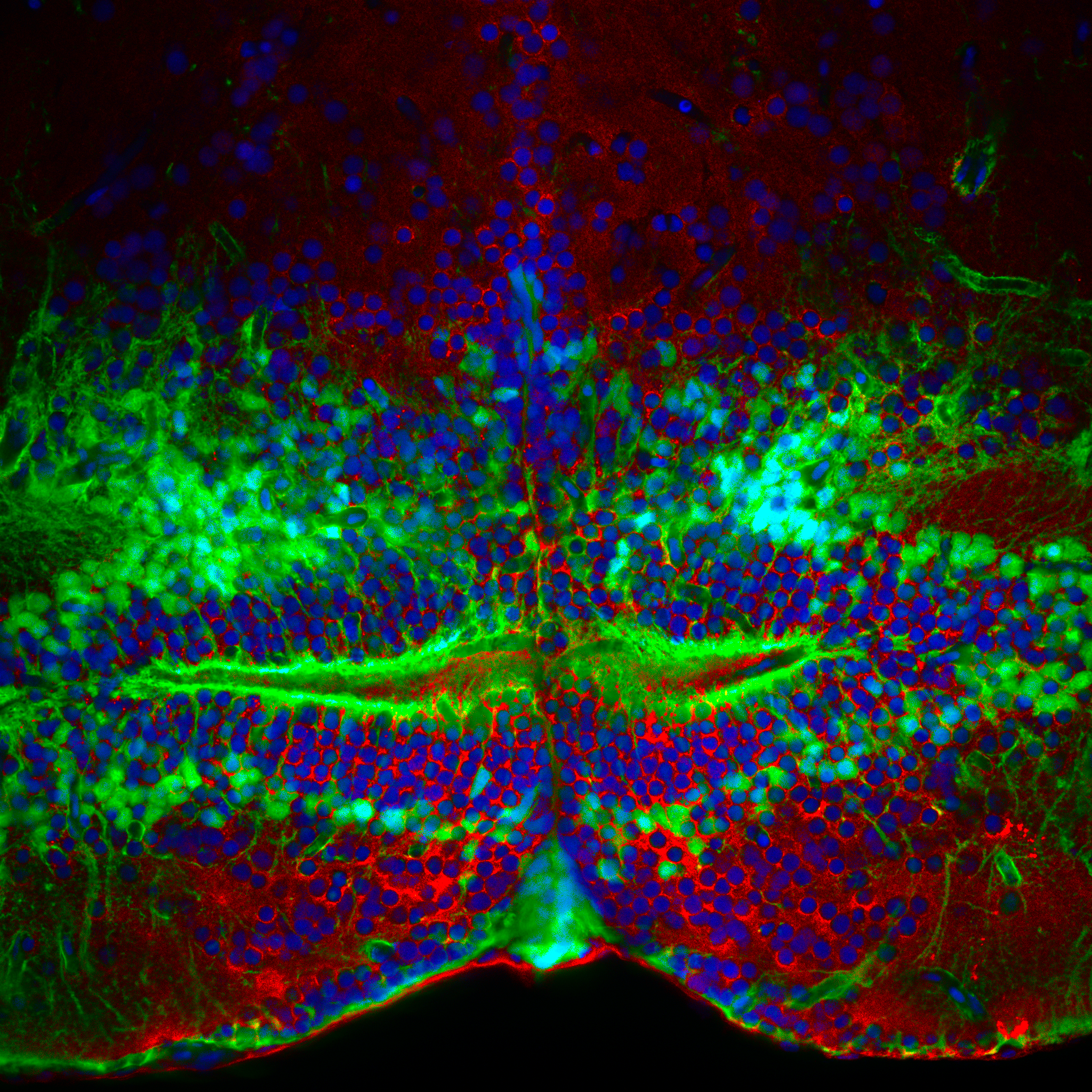
Photographer: Dr. Nicolas Diotel
Institution: Université de La Réunion, France
The Aromatase B-positive neural stem cells (green), the source of de novo brain estrogens, lie in a close and delicate association with serotonergic neurons (red) within the nucleus of the posterior hypothalamic recess of the adult zebrafish, while also gracefully lining the blood vessels. Their nuclei, stained with DAPI (blue), shimmer like distant constellations scattered across a nocturnal sky. Beyond the remarkable beauty of this cellular organization, poetry quietly finds its way into our minds, reminding us that even in the darkest of times, neuroendocrinology still rekindles our capacity to love. These magnificent stem cells unveil their most exquisite green attire, gathering before our eyes into the shape of a mouth with two slender lips, filled with the tenderness of serotonergic love. Slowly, delicately, they seem to draw near to us, offering the gentle kiss of a butterfly… fragile, silent, and infinitely soft… drifting into a sky of blue stars
🥈 Second Prize
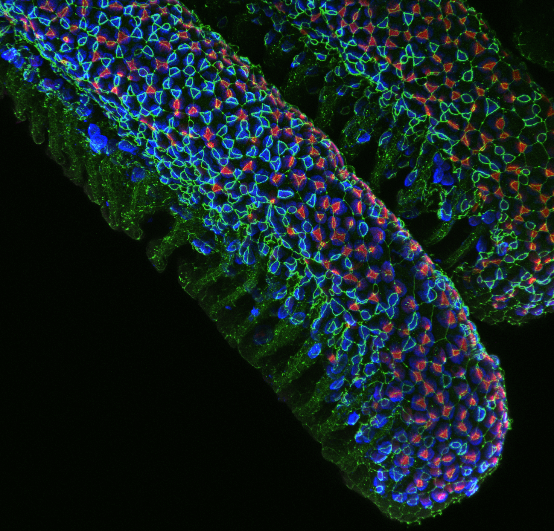
Photographer: Dr. Shang-Wu Shih
Institution: University of Michigan, USA
Prolactin-treated gills. Gill tissues from prolactin-treated medaka were fixed and immunostained to visualize branchial tight junctions and ionocytes, specialized epithelial cells that support ion transport, acid–base balance, and osmoregulation in fish. Prolactin is a key endocrine regulator of freshwater acclimation and promotes epithelial remodeling that helps maintain ion balance in hypoosmotic environments. This image highlights the organization of ionoregulatory cells and epithelial junctions in the gill, a central tissue for endocrine-controlled osmoregulation in teleost fish. The image was acquired using a ZEISS LSM 980 confocal microscope and processed in Fiji as a Z-stack projection.
🥉 Third Prize
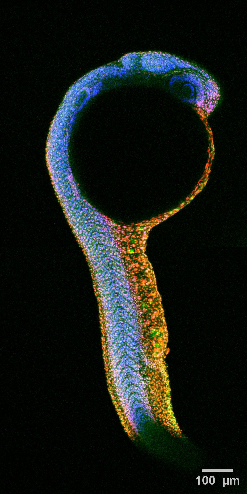
Photographer: Aurea Orozco’s laboratory
Institution: Universidad Nacional Autónoma de México, Mexico
Whole-mount immunolabelling of a 24 hour post-fertilization zebrafish (Danio rerio) embryo (lateral view). PCNApositive cells are shown in red, thyroid hormone receptor expresión is shown in green and nuclei were stained with DAPI (blue).
Thank You!
Congratulations to our three winners and thank you to every participant for sharing your work with the ISFE community. Your photographs beautifully demonstrate that science is not only about discovery—it is also about revealing the remarkable beauty of the natural world.
We also thank everyone who helped promote the contest by sharing our announcements and encouraging colleagues to participate.
We look forward to organizing future editions and continuing to celebrate fish endocrinology through science and photography.
The ISFE Executive Committee
